
Tretiakoff on Parkinsonism
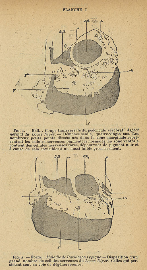 Plate 1, showing the appearances of the substantia nigra in a section of the brainThe Uzbek neuropathologist Konstantin Tretiakoff (1892-1958), who studied medicine in Paris, presented his MD thesis at a time when interest in parkinsonian syndromes had intensified as a result a result of the pandemic of encephalitis lethargica*. This epidemic commenced at the end of the First World War and led to a large increase in people with Parkinsonism.
In 1919 Tretiakoff reported research on the state of the substantia nigra (part of the mid-brain basal ganglia) in 54 human brains, six of which had belonged to people who had suffered from paralysis agitans and three from postencephalitic Parkinsonism.
In the six brains of those with paralysis agitans Tretiakoff noted a marked loss of pigmented neurones, together with swelling of the cell bodies of surviving neurones which he called ‘grumeaux’ degeneration (‘grumous’ meaning granular in microscopic appearance).
He found fewer pigmented nerves in the ‘zone ventrale du Locus Niger’ in these brains (see Figure 2, opposite) and in the brains from people with encephalitis lethargica (see Figure 6, below) than in brains from people free of these conditions (see Figure 1, opposite).
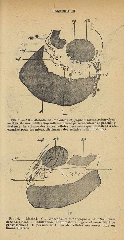 Plate 3, showing the appearances of the substantia nigra in a section of the brainAlthough not all neurologists accepted that the same underlying pathology was shared by the two conditions, Tretiakoff’s results extended the significance of Blocq and Marinesco’s findings which had pertained to a single case, and prepared the ground for further confirmatory studies.
*Encephalitis lethargica An inflammatory brain disease believed to be viral in origin which started towards the end of the First World War and became a global pandemic over the succeeding decade. With a mortality of some 40%, many survivors suffered profound alterations of consciousness, abnormal eye movements and Parkinsonism.
Planche I and III, reproduced above, compare the appearances of the substantia nigra (the horizontal swathe of white between the grey tissues) in a section of the brain of a woman who had developed dementia without signs of Parkinsonism in Figure 1, showing multiple black dots labelled ‘cn’ (representing ‘celles nerveuses pigmentées’) with the sparsity of such nerve cells in a section of the brain of a woman who had suffered from typical Parkinson’s disease in Figure 2 and a woman who had developed encephalitis lethargica in Figure 6.
Images in this section are courtesy of Bibliothèque interuniversitaire de santé (Paris).
In this exhibition
- James Parkinson's life and career
- Parkinson's London
- Parkinson's political activities
- Parkinson's fossils
- Tremors, agitations and palsy
- An essay on the shaking palsy
- Parkinson's neurological legacy: The 19th century
- Parkinson's neurological legacy: After Charcot
- Brissaud's lectures
- Problems in neurology
- Tretiakoff on Parkinsonism
- Awakenings
- Take me home
- Bibliography