
Hanson's preliminary work at King's, 1950-1953
 Jean Hanson in the laboratory Preparatory work required the isolation of suitable muscle fibres from a variety of sources in order to undertake comparison of structures.
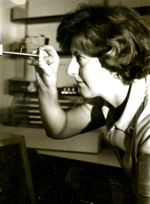
Hanson used established enzyme techniques such as those of Schick and Hass, and Szent-Györgyi, and teased out individual myofibrils to obtain rabbit, rat and frog muscle.
These could be subjected to study under the Unit's invaluable phase-contrast optical microscope that was especially useful in observing the banding of muscle fibres.
She introduced quantities of ATP (adenosine triphosphate) the energy-transferring molecule of living organisms, to view the consequent contraction of the fibres. More refined results were obtained by altering the concentration of ATP.
By measuring the changes in dark and light banding in samples under different conditions of contraction, conclusions might be deduced as to the structural relationships at work. Hanson published her preliminary findings in Nature in 1952.
Realising, however, that she lacked necessary expertise in electron microscopy necessary to produce comparative findings with the optical studies, she obtained a Rockefeller fellowship to work for a year between 1953 and 1954 at the Massachusetts Institute of Technology (MIT), already a pioneer in electron microscopy of muscle fibres under FO Schmitt.
In this exhibition
- South Africa in the nineteenth century
- Declaration of War 1899
- Arrival in South Africa
- On campaign
- The heat of battle
- Climate and landscape
- Peace: the Treaty of Vereeniging 1902
- The pioneering work of Professor Jean Hanson, 1919-1973
- Early career
- Biophysics at King's College
- Hanson's research on muscles
- What was known about muscles before Hanson?
- Why was the research important?
- Hanson's preliminary work at King's, 1950-1953
- Work with Dr Hugh Huxley
- The sliding filament hypothesis
- Hanson’s later career and legacy