
Discovering the structure of actin
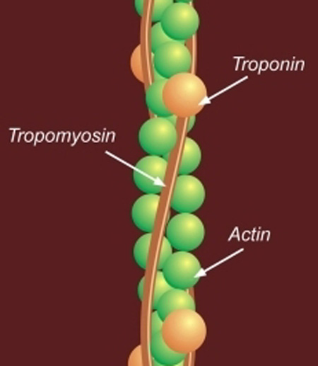 Diagram showing location of actin (1960s) During the 1960s, Hanson applied herself to discovering the actual structure of actin and thus obtain a clearer picture of how contraction occurs.
She and Lowy used the negative staining technique and the magnesium precipitation process to produce paracrystals of actin.
These revealed its essential helical structure and identified the vital role of tropomysin in the regulation of contraction. The results were also consistent with x-ray diffraction pictures.
This insight was an important component in the theory of cross bridges between the myosin and actin that provide the force for movement when the chemical energy conserved in ATP is converted into useful mechanical energy - a hypothesis developed out of the electron microscopy of Hugh and Andrew Huxley in the late 1950s.
Her work also helped explain the role of calcium uptake in muscle contraction.
Hanson's discovery at this time demonstrated how muscle protein fibres work at the molecular level to produce a more comprehensive and complete picture of muscle function and fully explain the microscope observations that underlay the sliding filament hypothesis.
In this exhibition
- South Africa in the nineteenth century
- Declaration of War 1899
- Arrival in South Africa
- On campaign
- The heat of battle
- Climate and landscape
- Peace: the Treaty of Vereeniging 1902
- The pioneering work of Professor Jean Hanson, 1919-1973
- Early career
- Biophysics at King's College
- Hanson's research on muscles
- Work with Dr Hugh Huxley
- The sliding filament hypothesis
- Hanson’s later career and legacy
- Applying the findings to all types of muscle
- Discovering the structure of actin
- The Muscle Biophysics Unit
- Hanson's Legacy