
The sliding filament hypothesis
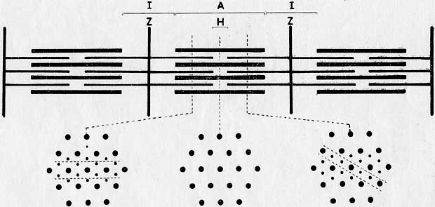 Diagram used to explain muscle fibre (1960s) The research also showed that individual protein filaments do not themselves undergo contraction but remain a constant length.
Instead, the filaments slide between one another and so collectively shorten or lengthen the whole muscle.
During muscle contraction, thin actin proteins interpolate between thicker myosin elements thus contracting the entire fibre and the muscle structure as a whole - the 'sliding filament mechanism'.
Furthermore, the simple elegance of the model was borne out by the visual evidence that A-bands always remained the same length apart, as would be expected if the model were correct.
The various sections of a muscle fibre are explained in a footnote in Randall’s memorial Emmeline Jean Hanson 1919-1973 in Biographical Memoirs of Fellows of the Royal Society 21 (Nov 1975), 312-344; 318:
Vertebrate striated muscles at rest-length have an extremely regular repeating system of transverse bands. Each repeating unit known as a sarcomere and commonly about 2.4 µm at rest length is bounded by two disks known as Z-lines. The dense band in the middle of the sarcomere is known as the A-band, is strongly anisotropic and about 1.6 µm long. In the centre of the A-band is a short zone of intermediate density and weaker birefringence; this is the H-zone. The I-bands are the zones (about 0.8 µm at rest length) of low density and lie on either side of the A-bands; each of them is bisected by a Z-line.
In this exhibition
- South Africa in the nineteenth century
- Declaration of War 1899
- Arrival in South Africa
- On campaign
- The heat of battle
- Climate and landscape
- Peace: the Treaty of Vereeniging 1902
- The pioneering work of Professor Jean Hanson, 1919-1973
- Early career
- Biophysics at King's College
- Hanson's research on muscles
- Work with Dr Hugh Huxley
- The sliding filament hypothesis
- Publication of the results in Nature, 1954
- The sliding filament hypothesis
- Hanson’s later career and legacy