
Signer's DNA
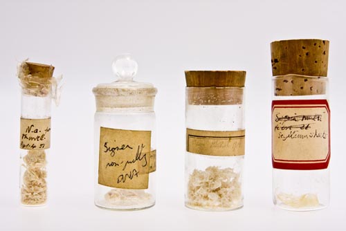 Bottles with Signer DNAIn 1950, Rudolf Signer, a Swiss biochemist based at the University of Berne, who had developed techniques of extracting high-quality DNA from cells, came to London to a Faraday Society meeting and distributed samples to research workers who expressed an interest, including Maurice Wilkins.
The DNA was extracted from a calf's thymus gland. Signer's DNA, and that supplied by Hans Schwander enabled experiments to be designed to explore the structure of this very large molecule.
The same year, Randall recruited Alexander Stokes. He was an experienced crystallographer and an excellent mathematician.
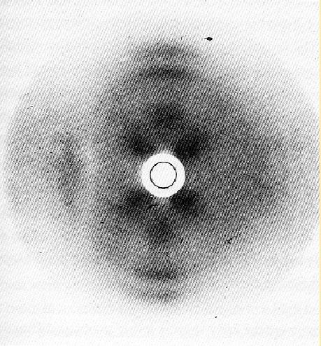 Gosling image of herring spermAlso in 1950, Raymond Gosling, a research student, began to study DNA in sperm heads which Randall was investigating using an electron microscope. Gosling used x-ray diffraction and was advised to pass x-rays through hydrogen to produce a more concentrated image than using air.
Wilkins found Signer DNA unique because it could be pulled or spun into thin filaments of remarkable uniformity that he felt might have a very regular structure and therefore give sharp x-ray diffraction patterns.
Wilkins was noted as being especially adroit at spinning out the fibres, and was described by Gosling as 'being like a wonderful spider'. (When Rosalind Franklin used similar DNA, she employed a highly polished and oiled microscope without the lenses to help pull the samples into the long strands necessary for successful photography.)
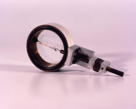 Sample holder used to photograph DNA at King'sThe fibres were so fine (10-30 μm) that they couldn’t be seen unless the light was arranged so that it could be reflected.
This not withstanding, Gosling used the samples to provide the first clearly crystalline x-ray diffraction picture of DNA prior to Franklin's arrival. He and Wilkins realised that precision could be improved by use of a microfocus x-ray tube attached to the camera.
One had just been developed by Werner Ehrenburg and W E Spear at Bernal's laboratory at Birkbeck College. Birkbeck gave the equipment to King's which was modified in King's well-equipped workshops.
In this exhibition
- Early work at King's
- Key individuals
- Key discoveries
- Further work at King's
- Background