
The momentum builds
 'B'-form of DNABy February 1951, Biophysics work at King's College London had grown to include 26 academic staff supported by a technical team of 23 led by Ted Benfield and Len Pitches that was the envy of many and was responsible for the motorised camera mounts and other bespoke precision engineering that often had to be built from scratch.
Gosling in particular remembers all new researchers being required to sketch out and explain precisely what they required, or thought they required, and then being led systematically through the practicalities of delivering a usable end product.
Linus Pauling, a structural chemist, published research in April 1951 on the structures in protein applying quantum mechanics to chemical bonding.
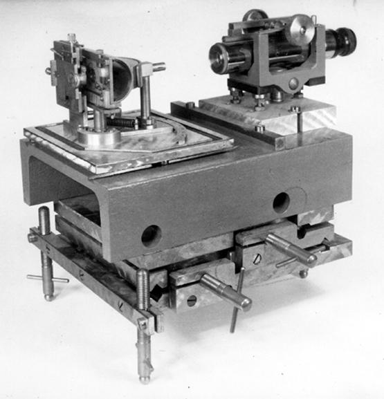 Tilting Micro-Camera and Stand Most significant among these was the proposition of the alpha helix, a basic structure present in many proteins.
King's scientists were interested but, like JD Bernal, puzzled by the fact the model had no way of calculating the x-ray diffraction and therefore no way of testing it.
At the request of Maurice Wilkins, Alexander Stokes devised a way of calculating diffraction from helices. Stokes used Bessel functions.
In May 1951, Wilkins lectured at an international conference at the Marine Biological Station at Naples and explained the reason for the concentration on nucleic acids at King's; the fact that when living matter is prepared in a crystal form the arrangement of molecules could be seen which in turn might lead to an understanding of the structure of the gene.
He illustrated the work with images unmatched in published literature.
 Workshop at King's College LondonBruce Fraser, physics research student in the King's Spectroscopy Group, was directed by Bill Price using Price's knowledge of chemical bonds and energies to build in late 1951 a helical model with phosphate on the outside but with three chains instead of two.
At King's it was thought that the density and water content indicated three chains (a view shared by Pauling and Astbury) but the model did not fit the x-ray images. Franklin argued modelling was premature.
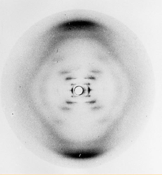
The real breakthrough came in May 1952, when Rosalind Franklin took a superb B form image, showing a clear X, which James Watson, in his account of the DNA discoveries, The double helix later described as the key image in recognising that DNA is a double helix, and has two chains.
In this exhibition
- Early work at King's
- Key individuals
- Key discoveries
- Further work at King's
- Background